
Mass Cytometry
Reagents for Mass Cytometry
Our magnetic nanoparticle reagents efficiently isolate your cells of interest
.Through funding by the National Science Foundation, we developed our magnetic quantum dot nanoparticles, known as the MagDot, by encapsulating both quantum dots and SPIONs within our patented polymer coating. When MagDots are conjugated to an antibody, they bind to cells of interest. One can magnetically collect the labelled cells and immediately analyze the cells fluorescently. Owing to its dual fluorescent and magnetic nature, there is no need to relabel cells fluorescently.

Incorporate MagDots and SpiDots in your Panel
Reagents
Design Your Panel
Applications
Applications
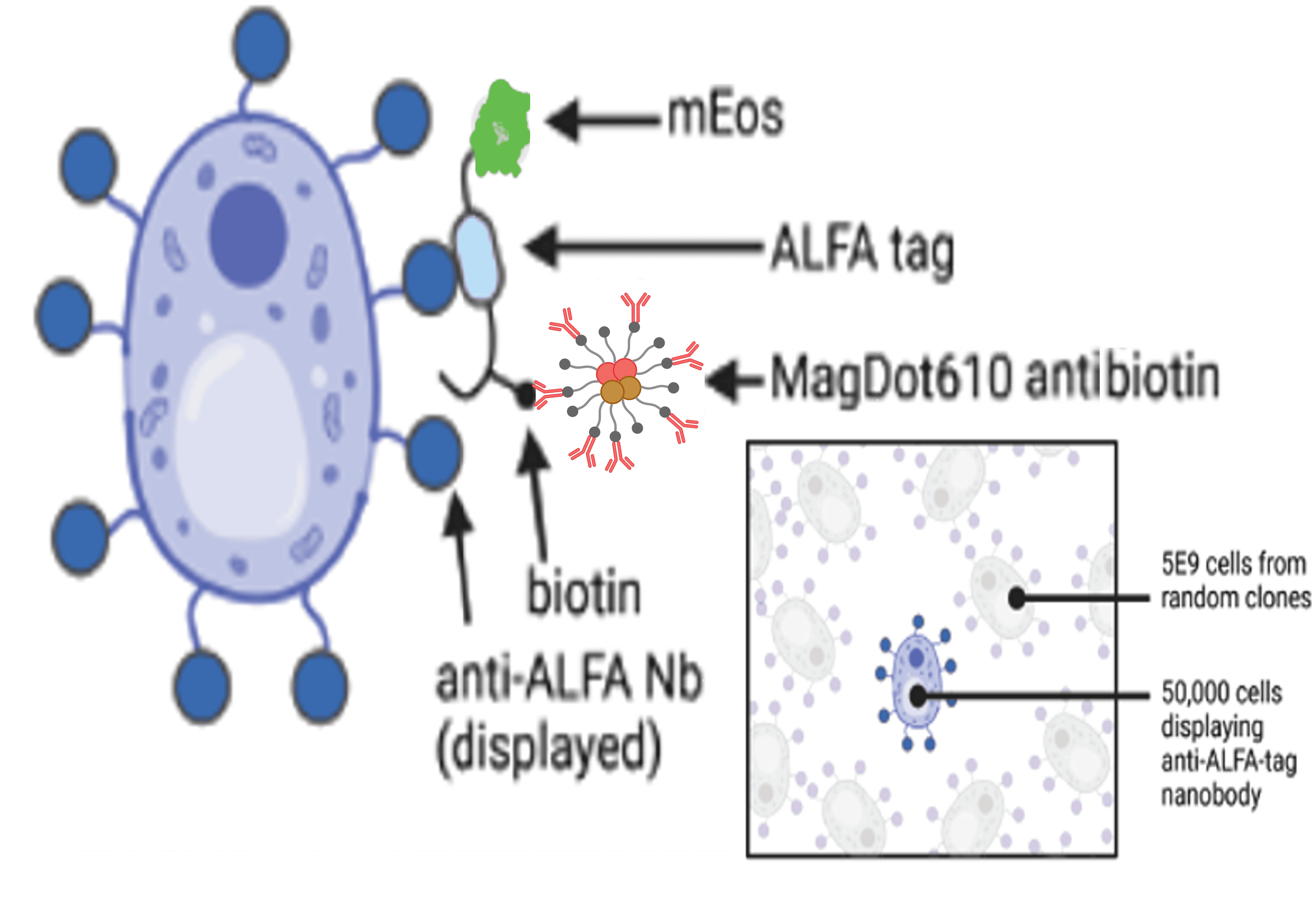
MagDot Antibiotin in Yeast Display Libraries
Yeast Surface Display is used for the expression and subsequent screening of desired proteins and peptides in the field of antibody discovery and development. Current techniques involved in separation are tedious and require multiple enrichment steps as well as individual labeling for each separation step to achieve the desired protein with sufficient purity. CQT’s MagDots that target biotinylated proteins allows for rapid and effective enrichment of antigen-binding clones in an open or column based magnetic separation system

T-Cell Isolation in an open magnet
Conventional T cell isolation involves MACs followed by using a fluorophore for FACs. With MagDots 610 antihuman CD4 one can isolate CD4+ T cells in a single step. Post enrichment in an open magnet, the cell populations tagged with the MagDot can be analyzed in a flow cytometer immediately, eliminating the need for a secondary reagent.

T-Cell Isolation in a packed column magnetic system
Conventional T cell isolation involves MACs followed by using a fluorophore for FACs. With MagDots 610 antihuman CD3 one can isolate CD3+ T cells in a single step. Post enrichment in a packed column magnetic system, the cell populations tagged with the MagDot can be analyzed in a flow cytometer immediately, eliminating the need for a secondary reagent.

MagDots for Enrichment of EGFR Positive Tumor Cells
Sponsored by the NSF, MagDot conjugated to EGFR were used to enrich cancer cells from a tumor biopsy. Heterogenous tissue samples have extreme difficulty being sorted on a flow cytometer, due to increased viscosity, high contamination debris and extracellular matrix, and rarity of cells to be enriched. EGFR positive cells in a cell suspension obtained from mechanically dissociated EGFR+ breast cancer biopsy, was labelled with MagDot 610 EGFR and collected in CoreMag2T for 30 minutes. Histogram B (in the middle) show the purified population with very few positive cells remaining in the non magnetic fraction.
Example protocol to enrich EGFR positive tumor cells with MagDots
